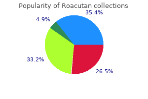
Roacutan"Purchase 10mg roacutan mastercard, acne extractor tool". By: Q. Mufassa, M.S., Ph.D. Associate Professor, Meharry Medical College School of Medicine The paper presents a study using image data from the Internet brain segmentation repository Segmentation strategies based on level sets are also commonly used for brain tumors acne mechanica discount roacutan 20mg overnight delivery. To overcome some of the challenges with setting up the speed function, a method that includes a dynamically updated speed function is presented in Taheri, Ong, and Chong (2009). Typically, level sets rely on boundary information for convergence, but an approach that includes both region and boundary information simultaneously. An illustrative example of an algorithm that presents a strategy for making use of multiple scans with varying spatial resolution is presented in Nie et al. The algorithm was applied to malignant glioma brain tumor segmentation and results were comparable to manually segmented tumor results by multiple subject matter experts. First, manual segmentation is prone to inconsistency, even when performed by experts. The second and more significant problem is that accurate "ground truth" data are often very hard to come by for many specific applications of interest. However, by selecting algorithms suitable to a specific problem, one can provide efficient complementary tools to assist clinicians in cumbersome activities such as locating and delineating the tissue regions present in the images. In the following, a variety of examples are provided to illustrate how region-, model-, or contour-based image processing algorithms can contribute in cancer treatment planning. One needs to (1) obtain the cranial and brain volume measurements, (2) localize gray and white and encephalic liquid as well, and (3) identify and localize potential lesions. Usually performed using standard parametric methods and Gaussian mixture models, the author identifies a limitation and lack of performance of those approaches in the case of an increasing number of parts including presence of abnormal tissues. Moving toward a nonparametric model, the author presents a Bayesian approach based on the Dirichlet mixture model. An important feature of this model is that inference on the number of components in the mixture is incorporated, a Markov random field provides some noise immunity as well. As expected, these methods performed well in segmenting abnormal tissues characterized by significantly different texture characteristics compared to the surroundings. However, the segmentation accuracy was only reported to be 75% at best (as compared to expert manual results), so it appears difficult to delineate precisely the entire tumor with these techniques. In this context, their method can at least trigger an alarm regarding the presence of a suspicious lesion, but cannot be considered as an accurate quantitative approach for the reported cases. The authors investigate the performance/time ratio of these automated techniques against manual delineation. They note performance results in terms of a volume overlap (a) (b) measurement, Vo, which is defined as the ratio of the intersection to the union of the automatic and manual segmentation volume results (optimal value is 1. Simultaneous segmentation of both cortical surfaces is performed using a coupled level-set approach. For example, for the automatic segmentation of the rectum on 44 total patients, 54% of the patient results were considered to be "acceptable," "good," or "excellent," while 45% were considered "not acceptable. Their approach is designed to be flexible so that segmentation results (details of which are left to the paper) can be easily refined by tweaking the input parameters. Quantitative assessment of the delineated area based on the Jaccard similarity metric (Js) (Jaccard 1901) provides information of the treatment progress. At first, the image is thresholded to extract the position of the liver, and then morphological operators are applied to extract the tumor candidates. Solomon, Butman, and Sood (2006) present the combination of a spatiotemporal model and a Markov model to perform fourdimensional brain tumor segmentation. Segmentation strategies range from data-driven (bottom-up) methods to model-driven (top-down) methods, and also include a host of techniques that are combinations of both (hybrid techniques). For cancer-specific applications, the most commonly researched and employed techniques include various types of pixel-based classification. Other approaches gaining popularity for brain segmentation are model-based strategies such as atlas-based segmentation. There is a wealth of research being published in each of these categories, and the purpose of this chapter was to introduce the reader to the most promising techniques so they can (1) choose a type of segmentation strategy most applicable to their cancer imaging research and (2) use the information as a starting point for implementing their own segmentation techniques that build upon the wealth of research being performed.
Cephalometric and histologic changes produced by extra-oral high pull traction to the maxilla of Macaca mulatta acne glycolic acid order roacutan with visa. Is longitudinal bone growth influenced by diurnal variation in the mitotic activity of chondrocytes of the growth plate Long-term mandibular adaptations to protrusive function: an experimental study in Macaca mulatta. Experimental and cybernetic approaches to the mechanism of action of functional appliances on mandibular growth. Cephalometric changes associated with treatment using the activator, the Frankel appliance, and the fixed appliance. The principle of the Andresen method of orthodontic treatment: a discussion based on cephalometric x-ray analysis of treated cases. Long-term effects of chincap therapy on skeletal profile in mandibular prognathism. Experimental and postexperimental response to anteriorly directed extraoral force in young Macaca nemestrina. Treatment response and long-term dentofacial adaptations to maxillary expansion and protraction. Skeletal response to maxillary protraction with and without maxillary expansion: a finite element study. The effects of maxillary protraction therapy with or without rapid palatal expansion: a prospective, randomized clinical trial. Timing for effective application of anteriorly directed orthopedic force to the maxilla. Quantitative analysis of the orthodontic and orthopedic effects of maxillary traction. Changes in facial dimensions associated with the use of forces to retract the maxilla. Long-term effect of treatment with the headgear-Herbst appliance in the early mixed dentition. The effects, limitations, and long-term dentofacial adaptations to treatment with the Herbst appliance. Prospective, multi-center study of the effectiveness of orthodontic/orthognathic surgery care in the United Kingdom. A roentgenocephalometric study of skeletal changes during and after chin cup treatment. Cephalometric variables predicting the long-term success or failure of combined rapid maxillary expansion and facial mask therapy. Midfacial protraction with skeletally anchored face mask therapy: a novel approach and preliminary results. Orthopedic traction of the maxilla with miniplates: a new perspective for treatment of midface deficiency. Threedimensional analysis of maxillary protraction with intermaxillary elastics to miniplates. Comparison of two protocols for maxillary protraction: bone anchors versus face mask with rapid maxillary expansion. Longitudinal changes in facial type in cases with vertical and horizontal mandibular growth directions. A cephalometric evaluation of anterior openbite correction with the magnetic active vertical corrector. Comparison of the effects of passive posterior bite-blocks with different construction bites on the craniofacial and dentoalveolar structures. Treatment effects of the bionator and high-pull facebow combination followed by fixed appliances in patients with increased vertical dimensions. Treatment of severe anterior open bite with skeletal anchorage in adults: comparison with orthognathic surgery outcomes. Craniofacial distraction with a modular internal distraction system: evolution of design and surgical techniques. Mandibular lengthening by distraction osteogenesis using osseointegrated implants and an intraoral device: a preliminary report. Stability of anterior openbite correction with multiloop edgewise archwire therapy: A cephalometric follow-up study. Furthermore, cephalometric analysis quantifies dentoskeletal relationships in angular and linear measures, which are not entirely representative of the threedimensioanl multidimensional interrelationship of craniofacial structures.
If the rectus abdominis is hypertonic and short it can be released and lengthened by laying supine over the curvature of a gym ball skin care trends purchase 30mg roacutan overnight delivery. The mobilization strap helps to apply the technique along the specific vector of resistance. A sustained Grade 4 mobilization technique is used to mobilize the adhered connective tissue within the fibrotic capsule. Find the greatest vector of resistance and explore all directions and limits of the functional range of motion. Ensure that the femoral head remains centered throughout the technique (palpate it anteriorly), and release any hypertonic muscles that arise at any time. Mobilization with movement (also known as a Mulligan mobilization or active release technique). After the passive technique has released the connective tissue, active mobilization with movement is useful. Maintain the lateral distraction of the hip along the line of the greatest vector of resistance and have the patient actively move further into, and out of, a variety of combined hip movements. If the technique has been successful, the femoral head will now be centered and remain so through an increased range of motion of the hip joint. Test the new functional range of hip motion (range where the femoral head remains centered) and repeat the mobilization technique, if necessary. Integrate the new functional range of motion into a home practice that has meaning for see them. Home practice for integrating hip mobility into tasks requiring extension will be covered later. With one hand, palpate the femoral head and support the lower extremity with the other. Take the hip joint to the motion barrier of flexion, adduction, and internal rotation (no pain should be provoked and the femoral head should remain centered in the acetabulum). Hold this femoral position and find the vector that provides the greatest resistance to lateral distraction by leaning back slightly into the mobilization strap. Once found, stop oscillating and sustain a strong, but painfree, lateral distraction force (Grade 4) in the line of this vector until you feel the connective tissue release. At home, instruct the patient to use either their hands or a towel to support the thigh and to then bring the knee towards the chest only until they feel a resistance to this motion (the first barrier). They can then apply a gentle hold/relax technique to have the hamstrings facilitate posterior rotation of the innominate. Weight bearing activities, such as walking, standing, and sitting, should be minimized during the first few days. For home practice, have the patient pull their knee towards their chest to posteriorly rotate the innominate relative to the sacrum. The innominate is often positioned in anterior rotation relative to the sacrum, and the joint does not unlock during vertical loading tasks. Active posterior rotation is reduced compared to the opposite side, as is the passive joint mobility. The neutral zone is reduced compared to the other side, and the end feel in the elastic zone of motion is firm. Once again, distraction of the joint according to specific vector analysis of the direction of greatest resistance is the mobilization technique of choice. With the other arm/hand support the femur, flex the hip, and posteriorly rotate the innominate relative to the sacrum until the motion barrier is perceived both with your posterior hand and with the flexing femur. If the hip can be taken into flexion/adduction/internal rotation with no impingement in the groin, the technique can proceed. Vary the direction of this force to find the specific vector of greatest resistance. The sacroiliac joint is capable of only a small amount of motion, and therefore it should be possible to restore all of its mobility in one treatment. Once this vector is found, a sustained Grade 4 mobilization technique is used to release the fibrotic connective tissue. There is rarely any need for a home practice to maintain the range of motion gained.
Syndromes
In vivo magnetic resonance spectroscopy: Basic methodology and clinical applications acne surgery purchase cheapest roacutan. Amide proton transfer imaging of 9L gliosarcoma and human glioblastoma xenografts. Using two chemical exchange saturation transfer magnetic resonance imaging contrast agents for molecular imaging studies. The magnetic resonance frequency 0 is linearly dependent on B 0 and on the gyromagnetic ratio of the nucleus, as 0 = B 0. The chemical shift is determined from the difference between the frequency of the peak of interest and the frequency of a standard compound, divided by the resonance frequency of the spectrometer. Since the numerator is typically in hertz and the denominator in megahertz, the chemical shift is expressed in parts per million, and is independent of the spectrometer magnetic field strength. Moreover, each peak is characterized by an area under the resonance signal, which is proportional to the number of nuclei. The line width of each peak is dependent not only on its intrinsic T2 but also on the magnetic field inhomogeneity as a consequence of the susceptibility of the tissue. Field homogeneity can be improved and optimized by shimming the local field, using shim coils. In vivo, it is difficult to achieve a comparable spectral resolution due to tissue microscopic (due to paramagnetic components) and macroscopic. In single-voxel techniques, three orthogonal intersecting slices are used to generate a voxel to minimize signal from regions outside of the volume of interest. However, because of the longer echo time, the technique is not suitable to detect metabolites with short T2 values, or complex J-coupling network patterns. The slice can be selected by subtracting one data set collected without inversion from one data set collected with inversion of the selected slice. Since eight acquisitions are required to accomplish the spatial localization, the sequence cannot be used for localized shimming. Variation between these data collections, for instance, due to movement, will degrade the quality of spectra. These localized spectra can be processed to obtain images from individual metabolites. Optimized spherical acquisition weighted k-space sampling can be used to further reduce imaging time. Metabolites such as lactate, total creatine, and total choline that have concentrations in the millimolar range are readily detectable. At shorter echo times, additional signals from short T2 metabolites, such as mobile lipids, glutamine, and glutamate are detected. Because of their high concentrations, the signals from water (~100 M) and lipid triglycerides (as high as ~10 M in some tissues such as breast) dominate unedited proton spectra. These dominant signals mask signals from other metabolites, and therefore water and lipid suppression methods are required. Another approach for water suppression is using band selective refocusing, where the signal from the water remains defocused resulting in loss of water signal [13,14]. The latter approach has the advantage of retaining signals of exchangeable protons in metabolites that can be suppressed by standard water suppression techniques. Although suppressing the water signal is required to detect metabolite signals, the water signal is useful as an internal concentration reference for absolute quantification of metabolites, as described in more details in paragraph 3. Prior to a standard localization sequence, additional chemical shift selective inversion pulses are either switched off or on to invert the metabolites signals upfield and downfield from water. To obtain the metabolite or the water signals, the two data sets are subtracted or added, allowing the simultaneous detection of signal from water and metabolites [16]. The origins of these lipids are still a matter of debate, and their role as biomarkers in tumor diagnosis and treatment monitoring is still being evaluated. Because these lipid signals dominate the proton spectrum, lipid suppression is required to detect signal from metabolites with chemical shifts close to the lipid signals such as lactate (1. Because of their short T2, mobile lipid signals decay faster than metabolite signals. Therefore, by using longer echo times that allow the lipid signal to decay, it is possible to reduce the lipid signal in proton spectra. Buy roacutan 30 mg on line. What our customers have to say about our clean skin care products for sensitive skin.
|