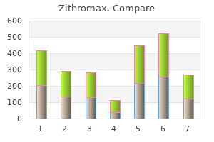
Zithromax"Zithromax 500 mg cheap, virus that causes hives". By: K. Bufford, MD Clinical Director, University of Florida College of Medicine Explicitly define each aspect of care that should be quantified to ensure a valid assessment of the most meaningful aspects of care antibiotics for uti azithromycin purchase zithromax 500 mg on-line. Review published literature (including guidelines and other performance measurement systems) with a team of clinicians and researchers with expertise in meta-analysis. For each measure, determine which data sources are available and define the data elements needed to construct it (including period of care). If chart abstraction is used, then interabstractor reliability needs to be measured; if patient survey is used, then item and unit nonresponse must be measured. For quality improvement, how many measures will be evaluated and/or whether a combined measure is necessary will need to be determined. Caution: To determine whether a provider has "improved" care over time or whether a provider is sufficiently different from others, a sample size calculation that incorporates the relevant statistical features of the "test" (within- and between-provider variability, size of test, significance of test) should be undertaken. Three phases are outlined in the development and implementation of performance measures: I. Essentially, the criteria require performance measures to be evidence-based, interpretable, actionable, well-constructed, and feasible. Table 5-4 is a summary of the established performance measures for chronic stable coronary artery disease. Acute Myocardial Infarction Coronary artery disease is the number one killer of men and women in the United States, accounting for 652,091 deaths in 2005. Although there has been decline in mortality through better utilization of the current guidelines, ongoing improvements in performance are anticipated to translate in to even greater reductions in mortality and morbidity. Measures made involving the initial hospitalization are typically made over the length of the hospitalization unless otherwise noted. All the performance measures discussed below are based on guidelines previously developed for treatment of a specific disease. Table 5-3 outlines the current status of available and developing performance measure sets for cardiology. For example, using Medicare database information, only half of patients at hospital discharge for an acute myocardial infarction have been counseled to stop smoking, only 79% are prescribed beta-blocker, and only 74% are prescribed an angiotensin-converting enzyme inhibitor. Atrial fibrillation can be isolated, recurrent (2 episodes), persistent or paroxysmal (self-terminates without cardioversion or pharmacologic treatment), longstanding (>1 year) or permanent (episodes last for >30 seconds at a time). The performance measurement set established for atrial fibrillation and atrial flutter, are summarized in Table 5-7. For the performance measures, atrial flutter is included with atrial fibrillation in terms of thromboembolic risk. Only people 18 years of age or older with atrial fibrillation or atrial flutter of nonvalvular cause are included in the guidelines. There has been a marked increase in incidence over the past two decades as the population ages. Chronic anticoagulation therapy with warfarin for patients with >1 moderate risk factors or any high risk factors. Risk factors for systemic embolism or ischemic stroke were determined from 5 primary prevention randomized trials with untreated control groups. These performance measures are derived from guidelines of the American College of Cardiology and American Heart Association, developed for patients >18 years of age evaluated in an office-based setting. The performance measures emphasize Class C and D heart failure as outlined in Table 5-10. Stages of heart failure used in the performance measure set are detailed in this table. Any high risk factor or more than 1 moderate risk factor *Adapted from Fuster et al. The period of assessment for inpatient performance measures is from admission to hospital discharge. Beta-blocker therapy is not included at present in the inpatient heart failure performance measure set, since it should be started when the heart failure patient is considered clinically stable. Given the complexities of assessing clinical stability through chart review, beta blocker utilization is not a performance measure at this time. Written instructions or educational materials are to be supplied to the patient or caregiver at the time of discharge from the hospital and should include: a follow-up appointment, dietary and activity restrictions or recommendations, regular assessment of weight, and a plan if symptoms of heart failure should recur or worsen. Suction catheters should not be secured to the chest wall to abrogate the potential for muscle injury and hemorrhage with removal antibiotic treatment for cellulitis order zithromax from india. Ideally, preservation of the long thoracic nerve ensures innervation of the serratus anterior. Innervation of the latissimus dorsi muscle is ensured with preservation of the thoracodorsal nerve that accompanies the neurovascular bundle of the posterior axillary space. Inset depicts position of closedsuction catheters (18 to 20 French) placed through separate stab wounds that enter the inferior margin of the flap at approximately the anterior axillary lines. Pectoralis major muscle (cut) Serratus anterior muscle Long thoracic nerve Axillary artery and vein Brachial plexus Subscapular artery Circumflex scapular artery Thoracodorsal artery, vein, and nerve Latissimus dorsi muscle Prior to flap closure, skin margins are carefully inspected to evaluate for devascularization that results from trauma of dissection or tangential incisions that contribute to skin necrosis and wound dehiscence. We prefer closure with interrupted 2-0 absorbable synthetic sutures placed in subcutaneous tissues with purchase of the cutis reticularis of the skin without tension. Optionally, the skin may be closed with subcuticular 4-0 synthetic absorbable sutures or stainless steel staples. Steri-strips are applied across (perpendicular to) the incision when subcuticular sutures are used in closure. After irrigation, the closed-suction catheters are thereafter connected and maintained on continuous low to moderate suction with large, plastic, vacuum reservoirs. We prefer to use light, bulky dressings applied with adherent sealed closure of the surgical incision. Some surgeons prefer compression dressings over flaps, and thus, wrapping of the chest wall with compression dressings. This practice may initiate central damage to the flaps with the potential of necrosis if undue pressure is applied with taping. Typically, wound catheters may be removed when drainage becomes predominantly serous and has decreased to a maximum of 20 to 25 cc during a 24-hour interval. Shoulder exercises are initiated on the day following removal of the drainage catheters. Alternative Closure of Flap Defects With use of the classical Halstedian operation and sacrifice of large volumes of skin centrally, alternative measures are essential to close the central defect. However, with the conventional cytoreductive chemotherapeutic neoadjuvant principles used today, such maneuvers are rarely necessary and flap closure can usually be accomplished as a primary procedure. Large defect that is expectant with creation of large skin flaps inclusive of the periphery of the breast for T3 and T4 lesions with fixation to the pectoralis major. The partial-thickness skin graft held in position with a compression stent created with cotton gauze mesh. Alternatively, compression foam mesh as the stent may be applied over the split-thickness skin graft. In the latter circumstance, the defect may be grafted with split-thickness skin grafts (0. To immobilize the skin graft and enhance the probability of adherence (with "graft take" to the chest wall), the partial-thickness graft is stented with bolsters. In such closures, catheters are not necessary when skin grafts are applied to the defect. The stents placed over the split-thickness skin graft are not removed until day 5 or 6 postoperatively, unless undue drainage (serum, blood, suppuration) from beneath the grafts is evident. This practice increases the probability of graft adherence, which is further enhanced when postoperative shoulder immobilization for large defects is ensured. When patients do not require postoperative irradiation following Halsted radical mastectomy, these patients may be offered immediate breast reconstruction following the radical procedure. Breast reconstruction allows for cosmetic enhancement of the patient with maintenance of form and functional restoration. However, with the presence of adverse biological features and the inexact probability of positive margins, the surgeon must defer the reconstruction. In addition, 68,348 patients evaluated between 1994 and 1995 had similar experience, in that this was an underutilized consideration. With the advancement and utility of myocutaneous reconstruction, with or without implants, the use of immediate reconstruction has increased between 1985 and 1995 from 3. These reports emphasize the importance of patient education as an essential component to address and provide proper patient consultation and information for options that include reconstruction risk, recurrence, and long-term morbidity. Each column represents the percentage of patients with that stage disease who had the operation listed in that row; that is, 22. Simultaneously was a pattern for decreasing use of the modified radical mastectomy also in favor of application of more conservative procedures with decrease from 63. Buy 250 mg zithromax amex. Immune System Part 1: Crash Course A&P #45.
Because of their unique properties antimicrobial underwear for men purchase zithromax 250 mg otc, the ion-exchange resins are generally used for taste masking, drug stabilization, tablet disintegration, and sustained release applications. Cationic or anionic drugs can be complexed in to the structure of an ion-exchange resin due to its ionic structure. The stability of the complex inside the mouth masks the taste of the drug since the drug will not be free to interact with taste buds. The drug will be released in the gastric medium once the complex becomes unstable. Certain drugs have a poor shelf life due to their instability against moisture, light, heat, and so on and so forth. Shelf stability and bioavailability of these drugs increase when formulated with ion-exchange resins. Although this polymer generally swells fast and to a high degree, its swelling properties are significantly affected by pH, the presence of salts, and the ionic strength of the aqueous solution. Nevertheless, the swelling pressure generated by this polymer is sufficient to disintegrate tablet dosage forms in an aqueous medium. These polymers can also provide a sustained or a zero-order release due to their high swelling capacity. Biodegradable Polymers Alternatively the drug can be released from a dosage form as a result of polymer erosion. Erosion occurs because of biodegradation or swelling and might happen within the bulk of the polymer or may be limited to the polymer surface at a time. Porous and nonporous platforms can provide bulk and surface erosion, respectively. Polymers with ester and amide functional groups are susceptible to a hydrolytic degradation in strong acidic and basic environment. When a polymer starts to erode from its surface, the drug imbedded within the polymer matrix will be released at a rate depending on the erosion rate of the polymer. If erosion happens in bulk, a much faster release is expected as an enormous number of hydrolysable sites are simultaneously cleaved up in water. Polyesters or copolyesters of lactic acid and glycolic acid, polycaprolactone, polyanhydrides, and polyethylene glycol are the most common synthetic biodegradable polymers, which are used for variety of pharmaceutical applications. Leuprolide (Eligard), a delivery system for prostate cancer, is supplied as an injectable suspension that utilizes the Atrigel technology for delivering the hormone leuprolide acetate. The delivery system consists of a biodegradable (lactide-co-glycolide) copolymer dissolved in a biocompatible solvent. The polymer gradually loses its organic solubility once it is injected subcutaneously. Doxycycline (Atridox), a bioabsorbable delivery system for the treatment of periodontal disease, also uses Atrigel technology to deliver an antibiotic, doxycycline hyclate. The matrix is made of nonbiodegradable but erodible polymers, which control the drug delivery due to its swelling. A polymer in its swollen form is mechanically weak and can be eroded at different rates depending on the swelling feature of the matrix. They are also increasingly used as taste-masking agent, stabilizer, and protective agent in oral drug delivery. Polymers can bind the particles of a solid dosage form and also change the flow properties of a liquid dosage form. Extensive applications of polymers in drug delivery have been realized because polymers offer unique properties which so far have not been attained by any other materials. Understanding Ion-Exchange Resins these are polymeric materials with two characteristics; they swell in an aqueous medium and they contain complexable and ionizable groups. This chapter provides basic concepts behind the behavior of polymers in the solid and solution states. Chapter begins with a general introduction on polymers and continues with different types of polymer structure and polymerization methods to make them. The major polymer concepts and properties, such as synthesis, topology, crystallinity, thermal transitions, molecular weight (averages and distribution), swelling, entanglement, rheology, and mechanical properties, are discussed in detail.
An introduction to the general topic of spectroscopy is presented in Chapter 4 and it is highly recommended that this section be reviewed before perusing this section antibiotics for uti at cvs 500mg zithromax fast delivery. This chapter is restricted to aspects of these techniques that are particularly relevant to biomolecules in a pharmaceutical context. Absorption spectroscopy in the ultraviolet and visible regions is a very versatile technique widely used with both proteins and nucleic acids. In contrast, in a diode array instrument, all wavelengths of light are put through the sample simultaneously and a spectrum created after absorption of the light. The resultant spectrum is then projected on to a diode array for detection purposes. Most importantly, by using mathematical fitting techniques to interpolate between the individual wavelengths detected by the diodes (typical spaced at 0. The high wavelength tail is often used, however, to detect proteins during various forms of chromatography. Phenylalanine (Phe) manifests a weak peak with marked vibrational structure between 250 and 270 nm, tyrosine (Tyr) a stronger, pH-dependent multicomponent peak from 250 to 290 nm, and tryptophan, the strongest absorbing side chain (another multicomponent peak) from 250 to 300 nm. In the case of proteins, the broad overlapping nature of these three contributions results in a broad peak centered between 277 and 287 nm (primarily from Trp and Tyr) with weak undulating bumps in the spectrum below 270 nm (due to the vibrational fine structure of the Phe contribution) and a marked shoulder for Trp at approximately 290 nm. Zero-order absorption spectra of four proteins, a peptide and a viral particle (upper panel). When their second derivative is calculated, however, usually six peaks are seen corresponding to the three classes of aromatic side chains (bottom panel). The important point here is that the position (wavelength) of each of these peaks is sensitive to the polarity of their environment. Thus, as the structure of a protein is altered, to the extent that one or more aromatic side chains experiences a subsequent change in its immediate environment, changes in the position of the derivative absorption peaks provide a measure of conformational change (and potentially physical degradation). The folding and unfolding of a protein as induced by temperature, pH, or a potential unfolding agent like urea, guanidinium hydrochloride, alcohols, chaotropic salts, and detergents can be simply followed by this method if their optical properties do not interfere with the measurements. If colors are then assigned to the major components, a map of temperature versus pH displays different colored regions corresponding to different physical states of the protein. These include metals such as copper or iron as well as lanthanides (with Tb and Co the most commonly employed), which can be used as calcium analogues. Fluorescence53,54 Probably the most versatile spectral technique for macromolecular structural analysis is fluorescence spectroscopy. When a chromophore such as the indole side chain of tryptophan in proteins is raised to an excited (singlet) state, rather than return to the ground state through internal conversion processes as in an absorbance measurement, it can do this by the emission of a photon. If this emission is from a triplet state, the process is known as phosphorescence (the combination of the two is called luminescence). For brevity, we will not be concerned with the latter here although the technique of phosphorescence can be quite useful in the analysis of proteins. In the case of fluorescence, the (relatively) long periods of time spent in the excited states (10-3 -10-9 s versus <10-15 s for absorbance) allow various types of interactions with this state. The spectrum of this emission is always at longer wavelengths than the absorption band(s) because prior to emission of fluorescence photons, energy is lost as the excited state returns to its lowest vibrational energy level. As a first approximation, the absorption and emission spectra are mirror images of one another. In addition to emission spectra, the lifetime of the excited state (; the time it takes fluorescence to fall to 1/e of its initial value) can also be measured. The amount of emission can be measured either in terms of its quantum yield (the number of photons emitted divided by the number absorbed) or by simple intensity changes at a fixed wavelength. Fluorescence is usually measured at right angles to the exciting light, with the emission monochromoter scanned. Alternatively, the excitation monochromoter can be varied and emission monitored at a fixed wavelength to produce a version of the absorption spectrum known as an excitation spectrum. Lifetimes are measured by either exciting with single pulses of light and measuring their emission decay or determining shifts in the phase of the emitted photons after modulation of the exciting light. At each T/pH condition, the protein is represented as a six-dimensional vector which is truncated to the three largest contributions. The regions with different shaded characteristics represent different structural states of the protein.
|