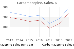
Carbamazepine"Discount 400mg carbamazepine with mastercard, muscle relaxant bruxism". By: B. Yorik, M.A., M.D., Ph.D. Clinical Director, Medical College of Georgia at Augusta University In that study muscle relaxant 750 mg trusted 100mg carbamazepine, depressed patients were randomly assigned to right unilateral versus bitemporal placement. Unlike previous studies, the investigators carefully quantified the electrical dose (charge in mc) of the electrical stimuli as described above. Utilizing a dosage titration procedure in which initial settings on the machine were at the lowest value, with progressively higher dosages used in one treatment session until a seizure ensued, they calculated precisely quantified seizure thresholds for each patient. At subsequent sessions, this value was also used, and if no seizure ensued, then somewhat higher values were used, so that along the course of treatments, stimulus dosages at or just above seizure threshold were used. Blindly evaluated depression ratings showed that remission rates were about twice as high with the bitemporal group as the unilateral group. Seizure durations were identical between the two groups, thus dispelling the long-held belief that seizure duration directly correlates with efficacy. The results led to the natural question about what would happen if higher-dose stimuli were used. Cognitive testing revealed an advantage in terms of anterograde and retrograde amnesia for the unilateral groups versus bitemporal, but the high dose unilateral group had more impairment than the low dose unilateral group. This was the first study to provide evidence that suprathreshold dosing can increase both efficacy and cognitive side effects. Cognitive testing revealed advantages in acute anterograde and retrograde amnesia for the 6. This showed both equal efficacy and better cognitive outcomes for a unilaterally treated group versus the bitemporal comparator. A weakness of the study is that the precise degree of suprathreshold dosing, that is, the degree to which the dose exceeded seizure threshold, was unknown. Thus, for each patient in the trial, the degree to which final dosing exceeded the initial threshold was known. Results showed a clear positive correlation between the degree to which dosing exceeded threshold and chance of antidepressant response as well as severity of cognitive side effects. Results showed equal efficacy but also equal cognitive impairment in this ultra-high dose unilateral condition. To summarize, several trials have demonstrated that the degree to which electrical dose exceeds initial seizure threshold directly correlates with antidepressant outcome and cognitive side effects with unilateral placement, with higher doses (in the range of six times threshold and higher) apparently equaling the efficacy of bitemporal placement. Regarding the latter, higher electrical doses beyond minimally suprathreshold appear in one trial (Sackeim et al. By the same token, cognitive side effects also appear to increase the higher the electrical dose exceeds threshold. Does the combination of stimulus parameters make a difference, independently of electrical dose, in determining efficacy and cognitive side effects? A goal of much research over the past few decades has been to elucidate a combination of stimulus parameters that is most efficient in inducing seizures that are therapeutic and also minimize cognitive side effects. Long ago it was established that brief pulse, square wave stimuli caused much less memory impairment than sinewave stimuli (Weiner et al. Ultimately, the gold standard study to pursue the question of stimulus parameter combinations would be to have multiple groups of patients with various combinations of stimulus parameters, meanwhile keeping the total dosage (charge) the same. The problem that becomes apparent is that if one wishes to focus on the relative importance of a single parameter, say pulse width, then a study would need to be designed in which there are groups for different pulse widths, say one with high pulse width and the other with low pulse width. However, at least one other parameter would need to be manipulated accordingly in order to keep the total electrical dosage the same. For example, stimulus frequency may be high in the "low pulse width" group and low in the "high pulse width group. In order to tease out the true influence of pulse width, we would need to design a study in which each of the pulse width groups (high and low) is further divided into other groups in which each of the remaining parameters is systematically varied so as to keep charge equal. If all of the low pulse width groups have superior outcomes to all of the high pulse width groups, with various other combinations of current, frequency, and train duration, then we could confidently conclude that pulse width affects outcomes independently of the other parameters. Each of these groups was subdivided into two other groups, one receiving "ultrabrief" pulse width stimuli of 0. Thus, there were four groups: right unilateral ultrabrief pulse, bitemporal ultrabrief pulse, right unilateral standard pulse, and bitemporal standard pulse. Outcome measures included initial stimulus titration thresholds as well as assessments of depression severity and in-depth cognitive batteries.
Amiodarone or an implantable cardioverterdefibrillator for congestive heart failure zoloft spasms best purchase for carbamazepine. Efficacy and safety of sildenafil citrate in men with erectile dysfunction and chronic heart failure. Patients with severe limitation of functional capacity have a high risk of hospitalization and death, independent of ejection fraction. If no reversible causes of disease progression are identified, patients should be evaluated for advanced therapies (cardiac transplantation or mechanical circulatory support), or if not eligible for such therapies, for palliative care and hospice referral. It is reasonable to consider reduction of dose or withdrawal of neurohormonal antagonist therapy in patients with advanced heart failure and intolerable side effects. It is reasonable to consider deactivation of implantable cardiovertor defibrillators in patients with advanced heart failure and estimated survival of less than one year. Vasodilators and positive inotropic agents may be used acutely and chronically to alleviate symptoms in this population as part of a comprehensive palliative care program. Clinical Assessment the characterization of advanced heart failure (Stage D) is primarily determined by the functional capacity of the patient rather than the left-ventricular ejection fraction. Patients with advanced heart failure have poor quality of life due to severely reduced functional capacity despite optimal medical and device therapy. Simple activities of daily living such as bathing and dressing can induce severe dyspnea, often require long, frequent rests to complete the task, and leave the patient feeling exhausted. Other distressing symptoms in this group included profound fatigue, sleep disturbance, restlessness, lethargy, poor concentration, 67 hair loss, alteration of taste sensation, sexual dysfunction, and anorexia with weight loss. Careful questioning about symptoms during specific activities is important, as many patients slowly curtail activities that produce dyspnea or fatigue without conscious acknowledgement of their change in lifestyle, and come to accept frequent dyspnea and fatigue with minimal exertion as a "normal" aspect of their heart condition. If a severe limitation in functional capacity is detected, the patient should be further questioned about day-to-day changes in the symptoms, since patterns in the daily severity of symptoms can sometimes help determine treatment strategies. Specific questions on sleep patterns and the physical symptoms that interrupt sleep may also be helpful to identify strategies to reduce fatigue. Specific information on patterns of food intake can identify strategies to improve nutrition and stamina. Depression, chronic pain, impaired concentration, and an uncomfortable sense of restlessness are common symptoms in this stage of the disease. Specific questions on these issues are useful to identify these conditions as potential targets for palliative therapy. On physical examination, patients with advanced heart failure often demonstrate cachexia (characterized by bitemporal wasting, wasting of the musculature of the shoulder girdle, and generalized muscle atrophy) and appear fatigued. In patients with advanced heart failure and reduced ejection fraction, the pulse pressure is often narrowed, with a thready pulse or pulsus alternans evident on palpation of the radial artery. Laboratory data will often demonstrate worsening pre-renal azotemia and hyponatremia. Anemia is also more common in patients with advanced heart failure, probably due to a combination of factors that reduce red cell production and increase hemodilution. Diuretic resistance is another clinical marker of low-cardiac-output syndrome in patients with other features of advanced heart failure. Diuretic resistance can be rapidly assessed by a spot sample for urinary sodium concentration one hour after a dose of intravenous loop diuretic. If the diuretic dose is already in excess of furosemide 80 mg (or its equivalent with other loop diuretics), other strategies including combination diuretic therapy (as discussed in chapters 8 and 9) or additional intravenous therapy to increase cardiac output and renal perfusion should be considered. For patients with heart failure and reduced ejection fraction, imaging may detect an increase in the left-ventricular end-diastolic dimension from prior studies, decrease in the left-ventricular ejection fraction from prior studies, and new or worsening mitral and tricuspid regurgitation. For patients with heart failure with preserved ejection fraction, subtle changes in left-ventricular size and left-ventricular ejection fraction from the patient baseline may be present, although typically not outside of the normal range. Many of the signs and symptoms of advanced heart failure are related to severe reductions in cardiac output reserve. Right-heart catheterization may be considered to confirm the clinical suspicion of reduced cardiac output, and also to directly measure cardiac filling pressures (to rule out volume depletion as a cause of the worsening symptoms). Estimated cardiac outputs derived from the Fick formula are more reliable than thermodilution cardiac output measurements in patients with advanced heart failure. Due to the invasive nature of this procedure, right-heart catheterization is not recommended routinely for all patients, but it should be performed in patients who may be candidates for mechanical circulatory support and/or cardiac transplantation as discussed below (age less than 75 years with no non-cardiac disabling or life-limiting conditions). It is recommended to obtain an electrocardiogram and chest radiograph in the evaluation of a patient with worsening symptoms.
If the vein slowly refills from below back spasms 24 weeks pregnant order carbamazepine with a mastercard, this observation is consistent with elevation of the central venous pressures. If the vein remains flat, the extended jugular vein is probably attributable to a venous valve. This can be confirmed by release of the superior finger with observation of a rapid descent of the column of blood toward the heart. This maneuver is not necessary if one can observe distinct cardiac wave pulsations in the external jugular veins. Such pulsations can only arise from the heart, so they indicate that the vein is filling due to elevated pressures in the jugular vein. Another useful technique in patients where the internal jugular venous pulsations are not easily visible due to body habitus is to carefully observe the head and neck region for other visible veins, often just superior to the sternal notch, or sometimes further posterior near the trapezius muscle insertion on the neck. In patients with severe elevations of jugular venous pressure (>20 cm), the pressure waves may not be easily visible since they are above the angle of the mandible. In these patients, venous pulsations can often be observed at the angle of the mandible along with a bobbing earlobe, or occasionally in veins visible over the temple or the forehead. If all of the above methods fail to provide an estimate of the jugular venous pressures, inspection of the veins in the upper extremity can provide a rough estimate. An important caveat is that upper-extremity veins may become obstructed due to thrombosis in patients with frequent phlebotomies or indwelling intravenous lines. In the absence of such obstruction, a distended upper-extremity vein without thrombosis will begin to empty when the mean level of the vein is greater than the central venous pressure. The patient should be examined in the standing position, starting with hands down by the side. The dorsal veins of the hand should be distended as the hand is well below the level of the heart in this position. If the central venous pressure is normal, the hand veins should start to collapse as the arm achieves a position horizontal to the ground at the level of the shoulder (since this position is above the level of the right atrium). If the hand veins do not collapse until the arm is raised above the shoulder, then it is likely that the central venous pressures are elevated. If the veins do not collapse after the arm is raised above the shoulder, the veins are likely obstructed and therefore will not provide an accurate estimation of central venous pressures. With practice, the assessment of jugular venous pressure can easily be accomplished at the bedside or office examination room in one or two minutes. Accurate estimation of the jugular venous pressure provides important information for the differential diagnosis of lower-extremity edema. Cardiac edema is invariably associated with elevation of jugular venous pressures, so the absence of elevated pressures strongly suggests a non-cardiac cause. Based on the physical examination of the edema, the estimated jugular venous pressures, and other pertinent details of the patient history, the further diagnostic evaluation for non-cardiac causes of edema can be individualized. Spot urine protein to creatinine ratio, serum albumin, and thyroid function tests are reasonable screening tests. For patients with strong suspicion of chronic venous stasis, referral for ultrasound imaging with a vascular specialist may confirm the diagnosis and also provide information relevant to treatment options. The presence of pulmonary rales has a broad differential diagnosis that should be considered at the time of initial presentation of suspected heart failure. The rales associated with heart failure are symmetrical and typically described as fine inspiratory crackles at the bases of the lungs. If the sounds are asymmetrical or extend to the apices, other primary pulmonary causes must be considered, including pneumonia, atelectasis, and interstitial lung disease. Suspicion of a non-cardiac cause of rales is heightened by absence of other findings of congestion on exam and a low brain natriuretic peptide level. In this setting, referral to a pulmonologist for further evaluation is reasonable. As mentioned above, many patients with heart failure may not manifest typical pulmonary rales, but rather may have diffusely decreased breath sounds with a bronchial quality.
For those patients who progressed during prehepatectomy chemotherapy muscle relaxant used during surgery carbamazepine 100 mg free shipping, 0% were alive at 5 years in comparison to 11% in responders. Therefore, surgery should only be considered in the setting of patients who have responded to preoperative chemotherapy or hormonal therapy, or both. Such cases of partial progression have the same median survival as patients who meet standard criteria for disease progression. The benefit of surgical resection in the group of patients with disease that is stable or responding to imatinib is not clear. Therefore, imatinib is accepted as the first-line treatment for metastatic disease. Disease progression is managed by dose escalation followed by second-line agents such as Despite heterogeneous selection criteria, 5-year survival rates fall into two groups. Several reports describe 5-year overall survival of approximately 25%;57,59 however, others report 5-year survival between 45% and 60%. Outcomes following hepatic resection may therefore merely reflect differences in tumour biology, or publication bias. Furthermore, 5-year disease-free survival rates are much lower than overall survival rates, suggesting that liver resection may function as a cytoreductive rather than curative procedure in these highly selected patients. Ovarian cancer Epithelial ovarian cancer represents the most common malignancy of the ovary, of which surgery and platinum-based chemotherapy remain the mainstay 137 Chapter 7 of treatment. The preliminary nature of these results precludes any definitive management recommendations. Similarly, survival following hepatectomy for metastatic disease is dependent on optimal cytoreduction, negative margin status, greater pelvic than abdominal disease and a longer recurrence-free interval. The study documented no operative mortality with 5-year disease-free and overall survival of 11% and 43%, respectively. Metachronous metastases and complete resection were highlighted as prognostic factors. In the study, 68 patients underwent surgery and were compared to a cohort of 20 patients who were eligible but refused an operation. Prognostic features included complete resection of liver lesions, negative margins, length of disease-free interval from resection of the primary and a leftsided primary lesion. Furthermore, an evidencebased approach to surgery combined with sunitinib or surafenib will hopefully be forthcoming. Ocular melanoma metastasises to the liver more frequently, but is more likely to be associated with isolated liver metastases than cutaneous melanoma. However, 75% of resected patients in this study developed recurrent disease, and the rate of recurrence was similar between the ocular and cutaneous groups. It seems reasonable to adopt a resectional approach in highly selected patients, i. This will occasionally lead to long-term survival, but patients with metastatic melanoma generally have a poor prognosis. Newer immunebased therapy combined with surgery may provide an added benefit in metastatic melanoma to the liver. Although biological agents such as interferon- and interleukin-2 have yielded promising response rates, these are rarely durable and are associated with significant toxicity. The available evidence for hepatectomy for metastatic melanoma is limited and consists largely of subset analyses from larger series of patients with non-colorectal liver metastases. A recent retrospective study evaluated all patients who presented with metastatic melanoma over the last decade at a single Australian institution. Overall 3-year survival was 40% with a median survival of 21 months, influenced largely by the number of metastases and the presence of multiple sites involved. Hepatic resection is controversial for these tumours and the available literature is scant. Metastatic oesophageal cancer is usually widely disseminated and is associated with a 5-year survival of 3͵% when multiple sites of disease are present and 7% when disease is limited to the liver. Both patients developed multiple liver metastases at 680 and 781 months postoperatively. Thus, although rarely feasible, hepatectomy followed by hepatic arterial chemotherapy may provide a limited survival benefit in chemosensitive oesophageal cancer with isolated liver metastases. Order carbamazepine 200 mg free shipping. Statin Side Effects.
|