Zymycin"Purchase line zymycin, antibiotics for dogs petsmart". By: Z. Faesul, M.B. B.CH., M.B.B.Ch., Ph.D. Deputy Director, Medical College of Wisconsin The normally marginated neutrophils do antibiotics for acne cause weight gain purchase zymycin 500mg otc, however, loosely adhere to the endothelium through the action of selectin and can be recruited very quickly. They are in dynamic equilibrium with the circulating pool, which is approximately equal to the size of the marginated pool. The size of the reserve pool in the bone marrow and in the vascular compartment depends on the rate of granulopoiesis, the life span of the neutrophils, and the rates of migration into the bloodstream and connective tissue. Transcription factors control the fate of hemopoietic cells, whereas cytokines and local mediators regulate all stages of hemopoiesis. Blood the nucleus of the band (stab) cell is elongated and of nearly uniform width, giving it a horseshoe-like appearance. Nuclear constrictions then develop in the band neutrophil and become more prominent until two to four nuclear lobes are recognized; the cell is then considered a mature neutrophil, also called a polymorphonuclear neutrophil or segmented neutrophil. Although the percentage of band cells in the circulation is almost always low (0% to 3%), it may increase in acute or chronic inflammation and infection. Signaling molecules from a variety of bone marrow cells initiate intracellular pathways that ultimately target a select group of synergistic and inhibitory proteins known as transcription factors. Times indicated along vertical lines are the approximate time between recognizable stages. M-1 wk indicates increase in number by mitosis for 1 week before differentiation begins. In addition to identifying the various intracellular transcription factors, recent studies have identified and begun to characterize numerous signaling molecules found in the bone marrow. These include glycoproteins that act as both circulating hormones and local mediators to regulate the progress of hemopoiesis and the rate of differentiation of other cell types (Table 10. Specific hormones such as erythropoietin or thrombopoietin, discussed in a previous section, regulate erythrocyte and thrombocyte development, respectively. Interleukins, produced by lymphocytes, act on other leukocytes and their progenitors. Any particular cytokine may act at one or more stages in hemopoiesis, affecting cell division, differentiation, or cell function. The isolation, characterization, manufacture, and clinical testing of cytokines (proteins and peptides that are signaling compounds) in the treatment of human disease are major activities of the rapidly growing biotechnology industry. Nearly all of them act on progenitor stem cells, lineage-restricted progenitor cells, committed cells, and maturing and mature cells. Therefore, the targets listed above are target lines rather than individual target cells. Although lymphocytes continuously proliferate in the peripheral lymphatic organs, the bone marrow remains the primary site of lymphopoiesis in humans. In mammals, these cells originate in bursa-equivalent organs such as the bone marrow, gut-associated lymphatic tissue, and spleen. The production and differentiation of lymphocytes are discussed in more detail in Chapter 14, Lymphatic System. The bone marrow sinusoids provide the barrier between the hemopoietic compartment and the peripheral circulation. In sections, the cells in hemopoietic compartment appear to lie in "cords" between sinusoids or between sinusoids and bone. It occupies the position normally occupied by a capillary; that is, it is interposed between arteries and veins. It is believed to be derived from vessels that have just nourished the cortical bone tissue. The sinusoid wall consists of an endothelial lining, a discontinuous basement membrane, and an incomplete covering of adventitial cells. The adventitial cell, also called a reticular cell, sends sheet-like extensions into the substance of the hemopoietic cords, which provide some support for the developing blood cells. They also play a role in stimulating the differentiation of developing progenitor cells into blood cells by secreting several cytokines. The lightly stained basophilic area reveals immature chondrocytes (arrows) within the perichondrium (P) infection 3 weeks after wisdom tooth extraction cheap zymycin on line. These cells are formative chondrocytes that are just beginning to , or will shortly, produce matrix material. In contrast, the nuclei near the bottom edge of the micrograph are fibroblast nuclei (Fib); they belong to the outer layer of the perichondrium. Note how attenuated their nuclei are compared with the formative chondroblast nuclei of the inner perichondrial layer. This cartilage is replaced by bone tissue except where one bone contacts another, as in a movable joint. In these locations, cartilage persists and covers the end of each bone as articular cartilage, providing a smooth, well-lubricated surface against which the end of one bone moves on the other in the joint. In addition, cartilage, being capable of interstitial growth, persists in weight-supporting bones and other long bones as a growth plate as long as growth in length occurs. The role of hyaline cartilage in bone growth is considered briefly below and in more detail in Plates 13 and 14. This section shows the cartilages that will ultimately become the bones of the foot. In several places, developing ligaments (L) can be seen where they join the cartilages. They are aligned in rows and are separated from other rows of fibroblasts by collagenous material. The hue and intensity of color of the cartilage matrix, except at the periphery, are due to the combined uptake of the H&E. The collagen of the matrix stains with eosin; however, the presence of sulfated glycosaminoglycans results in staining by hematoxylin. The matrix of cartilage that is about to be replaced by bone, such as that shown here, becomes impregnated with calcium salts, and the calcium is also receptive to staining with hematoxylin. The many enlarged lacunae (seen as light spaces within the matrix where the chondrocytes have fallen out of the lacunae) are due to hypertrophy of the chondrocytes, an event associated with calcification of the matrix. Thus, where these large lacunae are present, that is, in the center region of the cartilage, the matrix is heavily stained. It will constitute the synovial membrane in the adult and contribute to the formation of a lubricating fluid (synovial fluid) that is present in the joint cavity. Therefore, all the surfaces that will enclose the adult joint cavity are derived originally from the mesenchyme. Synovial fluid is a viscous substance containing, among other things, hyaluronan and glycosaminoglycans; it can be considered an exudate of interstitial fluid. The synovial fluid could be considered an extension of the extracellular matrix, as the joint cavity is not lined by an epithelium. The newly formed metaphyseal bone, which is admixed with this degenerating calcified cartilage and is difficult to define at this low magnification, has the same yellow-brown color as the diaphyseal bone. Later, the cartilage becomes calcified; bone is then produced and occupies the site of the resorbed cartilage. With the cessation of cartilage proliferation and its replacement by bone, growth of the bone stops, and only the cartilage at the articular surface remains. The details of this process are explained under endochondral bone formation (Plates 13 and 14). This photomicrograph shows a developing long bone of the finger and its articulation with the distal and proximal bones. Before the stage shown here, each bone consisted entirely of a hyaline cartilaginous structure similar to the cartilages seen in the figure above but shaped like the long bones into which they would develop. Here, only the ends, or epiphyses, of the bone remain as cartilage, the epiphyseal cartilage (C). It is found in the auricle of the external ear, in the auditory tube, in the epiglottis, and in part of the larynx. Zymycin 500mg with amex. Ethnobotany-Guided Identification of Antibiotic Resistance Modifying Agents.
This low-magnification photomicrograph of the spleen reveals the same components shown in the previous drawing virus protection program cost of zymycin. Note the capsule with several trabeculae projecting into the substance of the spleen. In the center, there is a trabecula containing a trabecular vein through which blood leaves the organ. The white pulp contains lymphatic tissue that follows and ensheathes the central artery. Splenic pulp, in turn, is divided into two functionally and morphologically different regions: white pulp and red pulp, based on the color of fresh sections. White pulp appears as circular or elongated whitish gray areas surrounded by red pulp. Branches of the splenic artery course through the capsule and trabeculae of the spleen and then enter the white pulp. Within the white pulp, the branch of the splenic artery is called the central artery. The nodules usually contain germinal centers, which, as in other lymphatic tissues, develop as B cells proliferate after their activation. In humans, germinal centers develop within 24 hours after antigen exposure and may become extremely large and visible with the naked eye. These enlarged nodules are called splenic nodules or Malpighian corpuscles (not to be confused with the renal corpuscles that have the same name). Lymph nodes enlarge because of edema and hyperplasia of lymphatic nodules and their cellular components. These include B lymphocytes, T lymphocytes, macrophages, and other antigen-presenting cells. In severe bacterial infections, lymphadenitis may be accompanied by lymphangitis, an inflammation of afferent lymphatic vessels that carry infected lymph into regional lymph nodes. Inflamed lymph vessels may be visible as red streaks under the skin in the affected area of lymphatic drainage. Common symptoms of acute lymphadenitis are swollen lymph nodes that are tender to palpation, fever, chills, loss of appetite, tachycardia, and general weakness. Lymph nodes are usually palpable and tender, with red discoloration on the overlying skin. In severe cases of suppurative necrosis (necrosis with pus formation), a fistula (false opening) may develop that allows pus to drain from the enlarged lymph node to the surface. The most common microbial organisms that cause lymphadenitis are streptococcal and staphylococcal bacteria. Other less common organisms are viruses (as in mononucleosis or rubella), protozoa, rickettsiae, fungi, and the tuberculosis bacilli. The majority of pale-staining cells within the germinal center are represented by B lymphocytes and macrophages; accumulation of T lymphocytes forms a distinct mantle zone that encircles the germinal center. In chronic lymphadenitis, lymph nodes are enlarged, but they are usually not tender. Red pulp has a red appearance in the fresh state as well as in histologic sections because it contains large numbers of red blood cells (Plate 40, page 484). Essentially, red pulp consists of splenic sinuses separated by splenic cords (cords of Billroth). Large numbers of erythrocytes, macrophages, lymphocytes, dendritic cells, plasma cells, and granulocytes reside within the reticular meshwork of splenic cords. The iron from destroyed red blood cells is used in the formation of new red blood cells; splenic macrophages begin the process of hemoglobin breakdown and iron reclamation. Megakaryocytes are also present in certain species, such as rodents and the cat, but not in humans except during fetal life. Specific features of the spleen in comparison to other major lymphatic organs are summarized in Table 14. The splenic or venous sinuses are special sinusoidal vessels lined by rod-shaped endothelial cells. The stratum basale is a single layer of small infection 6 weeks after c section purchase 250 mg zymycin with visa, mitotically active basal cells that are attached by hemidesmosomes to underlying connective tissue and by desmosomes to each other. The stratum spinosum contains several layers of larger keratinocytes that are attached to each other by desmosomes located at the ends of their cytoplasmic processes containing intermediate filaments (keratin filaments). The stratum granulosum is a distinct layer of flattened keratinocytes filled with keratohyalin granules (contain precursors to filaggrin, which aggregates keratin filaments and lamellar bodies containing lipids, which, when secreted, are responsible for the formation of the epidermal water barrier. The stratum corneum is the most superficial layer of terminally differentiated squamous cells (with no nuclei) that are entirely filled with keratin filaments. Melanocytes (5% of cells in epidermis) reside in the stratum basale and have long processes that extend between keratinocytes into the stratum spinosum. Melanocytes synthesize melanin pigment in melanosomes and during the process of pigment donation, melanocytes transfer them into adjacent keratinocytes. The reticular layer is deeper and is composed of dense irregular connective tissue containing type I collagen, elastic fibers, and larger blood vessels. Dermal papillae contain nerve endings and a network of blood and lymphatic capillaries. The hair follicle contains a reservoir of epidermal stem cells (follicular bulge) that are responsible for differentiation into hair-forming matrix cells. Hair is formed by the differentiation of matrix cells in the inferior segment of the hair follicle (bulb) to form the medulla, cortex (80% of hair mass), and cuticle of a hair shaft. Sebum is produced by holocrine secretion and is discharged via pilosebaceous canal into the hair follicle. Their secretory parts contain myoepithelial cells, the contraction of which is responsible for expression of sweat. They produce sweat that is similar in composition to an ultrafiltrate of blood in kidney. Nails are plates of keratinized cells resting on nail beds containing hard keratin that is formed in a nail root at the proximal part of the nail. As the nail plate grows, it moves over the nail bed with edges covered by skin folds. Under the dermis is a layer of loose connective tissue called the hypodermis, which is also generally referred to as the subcutaneous tissue or, by gross anatomists, as the superficial fascia. Typically, the hypodermis contains large amounts of adipose tissue, particularly in an adequately nourished individual. On the palms of the hands and soles of the feet, the epidermis has an outer keratinized layer that is substantially thicker than that over the other parts of the body. Accordingly, the skin over the palms and soles is referred to as thick skin, in contrast to the skin over other parts of the body, which is referred to as thin skin. In addition, the interface between the epidermis and the dermis is more complex in thick skin than in thin skin. The finger-like projections of the dermis into the base of the epidermis, the dermal papillae, are much longer and more closely spaced in thick skin. Although the layers of the epidermis are examined more advantageously at higher magnification, it is easy to see, even at this relatively low magnification, that about half of the thickness of the epidermis consists of a distinctive surface layer that stains more lightly than the remainder of the epidermis. The dome-shaped surface contours represent a cross-section through the minute ridges on the surface of thick skin that produce the characteristic fingerprints of an individual. One of the ducts is shown as it enters the epidermis at the bottom of an epithelial ridge. It will pass through the epidermis in a spiral course to open onto the skin surface. A sample of thin skin is shown here to compare with the thick skin in the above figure. Often, as in this tissue sample, the hair follicles and the glands, both sebaceous and sweat, extend beyond the dermis (De) and into the hypodermis. These processes meet with spinous processes of neighboring cells and, together, appear as intercellular bridges (arrows, inset). The keratinized cells are flat and generally adhere to other cells above and below without evidence of cell boundaries.
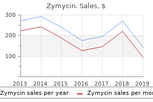
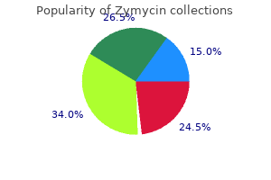
|