Procuta"Generic 40 mg procuta fast delivery, acne 30s female". By: R. Shakyor, M.S., Ph.D. Deputy Director, Mayo Clinic College of Medicine Intravascular ultrasound-guided direct intrahepatic portacaval shunt: midterm follow-up acne 7dpo 10mg procuta otc. Budd-Chiari syndrome: long term success via hepatic decompression using transjugular intrahepatic porto-systemic shunt. Endovascular approach to the trauma patient Uterine fibroid embolization: can fertility be preserved? Magdalena Jarzbek and Piotr Trojanowski Expert commentary Malgorzata Szczerbo-Trojanowska Case history A 20-year-old male arrived via ambulance at the A&E Department with severe epistaxis due to maxillofacial trauma following a fall from a height. Clinical tip In patients with massive epistaxis it is essential to localize the origin of the bleeding. Depending to the location of the source of bleeding, epistaxis is classified as anterior or posterior which require different methods of treatment. First-line therapies such as vasoconstriction, cautery, or anterior nasal packing are usually sufficient. Posterior bleeding, which occurs in the posterior superior part of the nasal cavity, is usually caused by major trauma and results from injury to vessels of larger calibre. Treatment involves posterior packing, surgical or endoscopic artery cauterization, or ligation. Intravascular embolization should also be taken into consideration as an alternative treatment [13]. This did not arrest the bleeding, and after 24 hours the haemoglobin level had dropped to 8. In addition to nasal packing the patient was treated with tranexamic acid and his hypertension was controlled. At the first attempt at removing the packing, bleeding in the posterior nasal cavity recommenced. The most common bony fracture after maxillofacial injury is fracture of the nasal bone [4]. Nevertheless, even in post-traumatic epistaxis it is necessary to exclude other possible causes or predisposing conditions, with injury triggering the bleeding. Medical history is important in making (continued) Case 16 Epistaxis: which embolic materials to use? In older patients the main causes of nasal bleeds are hypertension and anticoagulation treatment, whereas in younger patients trauma and malignancy are more common (see Table 16. Bleeding was still present at removal of the nasal packing so an endoscopy was performed. The patient was then consented for treatment with endovascular arterial embolization. These branches supply the inferior, middle, and superior turbinates, as well as the nasal septum. Arterial puncture under local anaesthesia and 144 Interventional radiology and endovascular procedures conscious sedation was performed. The nasal packing was then removed and there was no bleeding from the nose, indicating that cessation of epistaxis was achieved. He was advised to avoid physically strenuous work for a week and to strictly control his blood pressure. Discussion Epistaxis is a common problem with a 60% prevalence of at least one episode a lifetime in the adult population. However, only about 5% originates from the posterior part of the nasal cavity where it is considered to be a major medical problem and first-line treatments such as nasal packing, balloon, or systemic therapy often fail [3]. In post-traumatic patients the injury of vessels, pseudoaneurysm or arteriovenous fistula formation are likely to be the cause of the symptom. Embolization for bleeding has been a well-recognized procedure for at least three decades. It was first used for epistaxis in the 1970s and is currently a standard procedure when surgery is unsuccessful, contraindicated, or in systemic diseases with multiple bleeding sites [9,10]. Despite increasing acceptance of embolization in head and neck vascular diseases, considering it as an alternative to surgical or endoscopic ligation in epistaxis still remains controversial. One of the largest studies, conducted by Tseng et al [11], reviewed 107 patients with refractory epistaxis treated with intravascular embolization: 87% of this group suffered idiopathic epistaxis and in the remainder bleeding was post-traumatic or post-surgical. Examination Site A proportion of these lesions are usually sexually transmitted and are normally sited on the tip of the tongue or soft palate skin care in your 20s procuta 5 mg visa. Surface Papillomas have an irregular granular surface similar to the much large finger wart. Relations Papillomas are pedunculated in shape, arising from a narrow, stalk-like base. It can have a variety of clinical appearances and has many causes (see Revision panel 11. Movements of the tongue and cheeks are painful, and individual ulcers if present are very painful. In such circumstances, glossitis and oral manifestations are accompanied by signs and symptoms of the parent condition. It is important to obtain full medical history as stomatitis, and especially oral ulcers, may be a manifestation of an underlying metabolic disorder or be drug induced (nicorandil, antibiotics, chemotherapy). Candidiasis this is the result of monilial overgrowth (of Candida albicans) and has four forms in the mouth: Examination the physical appearances vary according to the cause. Aphthous stomatitis the cause is unknown but is thought to be an inappropriate and excessive immune response to a minor stimulus. Infection of the alimentary tract is common in children and in people with debilitating disease. It is well recognized as a complication of any antibiotic therapy that changes the balance of the bacteria in the alimentary canal. Recurrent aphthous ulcers are common in otherwise healthy young people, particularly at times of stress. The colonies of monilia on the tongue and mucous membranes look like patches of cream-coloured paint. The creases get soiled with saliva, and the moist environment encourages maceration of the epithelium and superinfection with commensal organisms (staphylococci and Candida). Angular stomatitis may be a manifestation of iron or vitamin deficiency, especially when accompanied by glossitis and oral ulcers. The attached gingiva around the teeth is bright red, and the pathognomonic feature is necrosis of the interdental papillae. The condition may be secondary to very poor oral hygiene or ushered in by a period of immunosuppression. Everything changed with antibiotics, and the disease, with its signs and symptoms, has now been all but eradicated in Western countries. Mucous patches and snail-track ulcers are part of the skin abnormalities that develop with secondary syphilis (see Chapter 4). Gummas can arise in the tongue or hard palate as painless masses that may ulcerate. Gangrenous stomatitis (Cancrum oris) the last reported case of gangrenous stomatitis in Europe was at the end of the Second World War. Now it is largely confined to a latitude range that includes dry barren sub-Saharan Africa. It normally commences as a small ulcer in the cheek that quickly extends with obvious signs of infection (redness, oedema, pain and loss of function). The dead tissue eventually separates, and with this the contours of the mouth and face are destroyed. It is estimated that 2 per cent of the population have a white patch in their mouth, but in only 2 per cent of this population is the condition premalignant. High-risk regions for malignant transformation are the ventrolateral surface of the tongue and the soft palate. Colour the lesions are (by definition) white, but there might be an erythematous component (erythroplakia). Shape and surface Leukoplakia can be homogenous (uniform) or heterogenous (verrucous, nodular or speckled). Lymph drainage the local lymph glands should not be enlarged, and pathological lymphadenopathy suggests a carcinoma. The tissues are friable so are vulnerable to trauma, and small superficial ulcers are a common feature of the condition. Generic procuta 20 mg with visa. ACNE SKINCARE ROUTINE | ACCUTANE | UPDATED | Conagh Kathleen.
They transferred me to the regional vascular centre skin care routine for acne buy online procuta, where the team decided that because this dissection was chronic and I was otherwise well, I did not need further intervention. I did well and was then discharged home but was readmitted a week later because I had recurrent chest pain and was coughing blood. A computed tomography scan found no clot in my lungs but showed that the aorta had increased in diameter from 5. I am still an inpatient at the regional vascular unit and am awaiting further surgery on my aorta. The surgeons have said that because of the complexity of my disease, I may be better suited to open rather than keyhole surgery. Is medical therapy still the optimal treatment strategy for patients with acute type B aortic dissections? Therapeutic management of patients with Marfan syndrome: focus on cardiovascular involvement. Whereas cardiopulmonary bypass facilitates open heart surgery for a number of hours, extracorporeal life support maintains tissue oxygenation for days to weeks in patients with life threatening respiratory or cardiac failure (or both). As technology advances, indications increase, and the numbers of specialist centres rise, more doctors are likely to find themselves assessing patients for early referral, discussing this support option with relatives, directly or indirectly managing patients on extracorporeal life support, and providing follow-up outpatient and community based care. During the recent H1N1 influenza A pandemic, one third of patients admitted to the intensive care unit with severe respiratory failure required extracorporeal life support. We used the search terms "extracorporeal membrane oxygenation", "extracorporeal circulation", and "extracorporeal life support". We also consulted the registry database of the Extracorporeal Life Support Organization, personal databases, reference collections, and contemporary textbooks. Adequate oxygen delivery to the tissue, therefore, requires a sufficiently high cardiac output and a haematocrit above 40%. The circuit consists of tubing taking deoxygenated blood from the patient, a pump, an artificial lung, a heat exchanger, and tubing returning oxygenated blood to the patient (fig 1). Venous-venous cannulation is used for isolated respiratory failure (tissue hypoxia secondary to hypoxaemia), whereas venous-arterial cannulation is used for cardiac failure (tissue hypoxia secondary to hypoperfusion) with or without respiratory failure (table 1). Deoxygenated blood flows from the venae cavae and oxygenated blood is returned to the right atrium (fig 1). Blood is removed distal to the right atrium and returned directly into the right atrium in an attempt to reduce mixing of deoxygenated with oxygenated blood, and to reduce recirculation of oxygenated blood within the circuit. It is impossible to prevent all mixing and recirculation, and Venous-arterial Cannulas are placed in a major artery and one or more major veins. Venous blood is oxygenated and pumped back directly into the arterial circulation, bypassing both the heart and lungs. Femoral arterial cannulation is common in adults (fig 2B), whereas the carotid artery is commonly cannulated in infants (fig 2C). In femoral artery cannulation a distal down flow cannula may be needed to prevent leg ischaemia, whereas in carotid cannulation, collateral circulation must be relied on for adequate brain perfusion. Existing transthoracic cardiopulmonary bypass cannulas may be used where extracorporeal life support is needed immediately after open heart surgery (fig 2D). In the presence of native heart function, oxygenated blood may not reach the proximal aorta, and this results in cardiac and upper body hypoxaemia (fig 2B). An increase in blood flow rate, a change to venous-venous extracorporeal life support, or placement of an extravenous return cannula may be needed. Anticoagulation Because of blood-surface interaction, an infusion of unfractionated heparin is necessary to prevent thrombosis within the circuit and embolism to the patient. Ventilation Positive end expiratory pressure is applied to the lungs and the ventilator is set to deliver low tidal volumes, low inspiratory pressures, and a low inspired oxygen fraction. These so called rest settings help to prevent ventilator induced lung injury, oxygen toxicity, and ventilator associated haemodynamic compromise. Extracorporeal life support is a support modality rather than a treatment in itself. It is invasive, complex, resource intensive, and can be associated with serious complications. Extracorporeal life support is, therefore, mostly reserved for patients with a high risk of death who have failed conventional management and where the underlying respiratory or cardiac disease is reversible. The greatest number of cases and highest survival rates have been reported in neonates with respiratory failure (table 2). Most of the evidence supporting extracorporeal life support for other age groups and indications consists of case reports, case series, and analyses of the Extracorporeal Life Support Organization database, although some randomised controlled trials have been reported.
She was reviewed at a vascular surgery outpatient clinic four weeks after the procedure and confirmed that her rest pain had improved acne under a microscope buy procuta pills in toronto. Emboli can be seen at the anterior tibial artery origin and the distal tibioperoneal trunk. Case 6 Iliac artery: surgical bypass, covered and uncovered stents 55 Discussion Most cases referred for endovascular treatment have diagnostic imaging confirming disease that is clearly attributable to their presentation. However, as in this case, the operator can be faced with partly non-diagnostic images, requiring an initial comprehensive catheter angiogram before forming a treatment strategy. Following the initial aorto-iliac angiogram showing a modest left-sided iliac stenosis, infrainguinal disease was suspected to account for the rest pain. The integrity of the infra-inguinal run-off is probably the most important independent predictor of stent durability in the iliac arteries [7,8]. Indeed, our case was not a high-risk surgical candidate and could equally have been offered open repair. Evidence base Surgery versus endovascular treatment Various review articles and meta-analyses have addressed the longevity of surgical bypass grafts versus endovascular treatment. These figures reflect advances in stent technology as well as operator technique, resulting in a general trend away from open repair of iliac occlusive disease. Morbidity and mortality also have to be considered when comparing surgical and endovascular treatments. Procedure-related mortality for endovascular intervention is less than 1% [15], and major complications are around 5% [16]. We acknowledge that potential differences in patient selection, and differences in potential morbidity outcomes, make a direct comparison of these outcomes difficult. In high-risk patients, extra-anatomical bypass (axillofemoral and femorofemoral) avoids the need for an open abdominal procedure. However, these procedures have inferior long-term patency compared with endovascular treatment [13]. This method has come under some criticism because it is not patient centred, with no reflection on the symptomatic success of procedures [17]. Whilst the results did not show a significant difference in long-term patency or re-intervention rates, the patients treated with angioplasty and selective stenting had a clinically better outcome. It also found considerable cost savings, as only 40% of the patients actually needed stenting, and thus concluded that selective stent placement should be the treatment of choice for iliac lesions. Interestingly, in the small group of iliac occlusions in the Dutch trial, 10 of 12 patients initially treated with angioplasty alone eventually required stent placement. In our practice, we preferentially adopt primary stenting after recanalization of occluded iliac segments. Contrary to the other findings in the Dutch trial, the complication rate in the angioplasty with provisional stenting was higher than in the primary stenting group (7% versus 4%) because of the complications of the initial angioplasty. In addition, primary stenting has been found to have a higher technical success rate [9]. Furthermore, the technical success of recanalization of an iliac occlusion seems to be higher with an antegrade approach. This study also found the major complication rate to be higher with the retrograde approach. As described by Bolia and Fishwick [4], a guidewire easily makes a dissection from the ipsilateral side, but re-entry is difficult closer to the aorta because the intima becomes thicker. Therefore this can lead to subintimal stent placement, which has a high stent thrombosis rate. A strategy of provisional angioplasty followed by pressure gradient measurement across the lesion (with a gradient of >10mmHg requiring a stent) is often awkward and time consuming, but is probably underutilized in our, and many other, practices and would potentially avoid unnecessary stenting in stenotic disease. Expert comment Imaging of lower limb vascular disease has been revolutionized by non-invasive imaging. However, there are potential limitations including signal drop-out from stents and other metallic structures such as clips, hip prostheses, etc. In reality, there is little that can be done from a technical perspective to reduce metallic artefacts from the metal alloys currently used in stents, and an alternative form of imaging may be required. It is certainly possible to significantly improve the quality of run-off imaging and reduce venous contamination with the use of blood pool agents and a technique of steady stateextended phase imaging [24]. Treatment of inflow disease may be enough to alleviate symptoms or heal ulceration, or may permit further infrainguinal intervention.
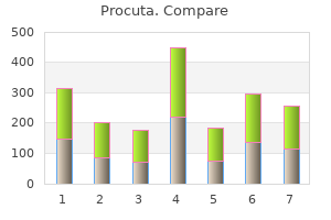
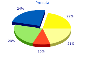
|